3. The Function of Nervous Tissue
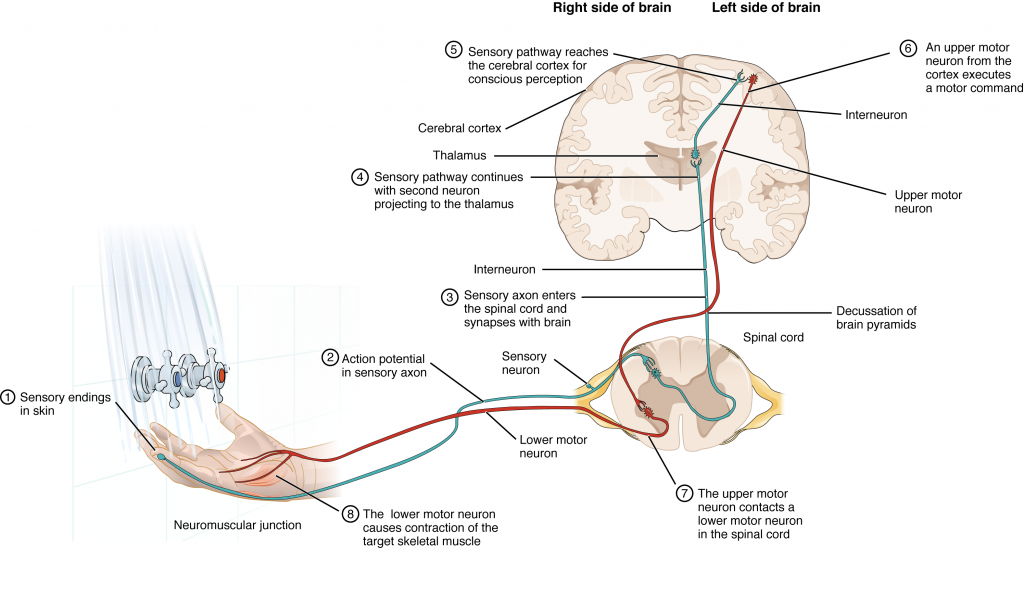
Having looked at the components of nervous tissue, and the basic anatomy of the nervous system, next comes an understanding of how nervous tissue is capable of communicating within the nervous system. Before getting to the nuts and bolts of how this works, an illustration of how the components come together will be helpful. An example is summarized in Figure 3.1.
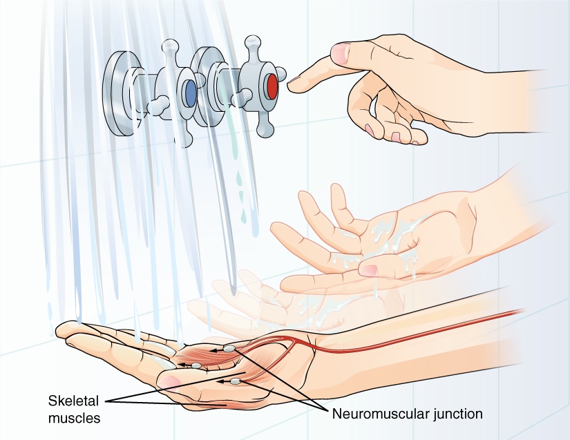
Imagine you are about to take a shower in the morning before going to school. You have turned on the faucet to start the water as you prepare to get in the shower. You put your hand out into the spray of water to test the temperature. What happens next depends on how your nervous system interacts with the stimulus of the water temperature and what you do in response to that stimulus.
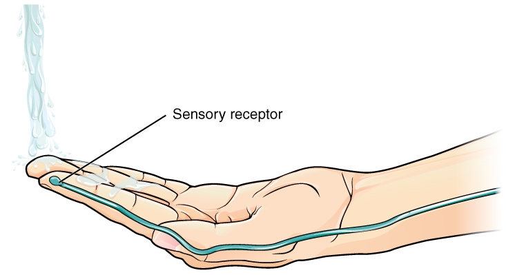
Found in the skin is a type of sensory receptor that is sensitive to temperature, called a thermoreceptor. When you place your hand under the shower (Figure 3.1 and Figure 3.2), the cell membrane of the thermoreceptors changes its electrical state (voltage). The amount of change is dependent on the strength of the stimulus (in this example, how hot the water is). This is called a graded potential. If the stimulus is strong, the voltage of the cell membrane will change enough to generate an electrical signal that will travel down the axon. The voltage at which such a signal is generated is called the threshold, and the resulting electrical signal is called an action potential. In this example, the action potential travels—a process known as propagation—along the axon from the initial segment found near the receptor to the axon terminals and into the synaptic end bulbs in the central nervous system (Figure 3.1). When this signal reaches the end bulbs, it causes the release of a signaling molecule called a neurotransmitter.
In the central nervous system (in this case, the spinal cord), the neurotransmitter diffuses across the short distance of the synapse and binds to a receptor protein of the target neuron. When the neurotransmitter binds to the receptor, the cell membrane of the target neuron changes its electrical state and a new graded potential begins. If that graded potential is strong enough to reach threshold, the second neuron generates an action potential at its initial segment f that graded potential is strong enough to reach threshold, the second neuron generates an action potential at its initial segment (Figure 3.1). The target of this neuron is another neuron in the thalamus of the brain, the part of the CNS that acts as a relay for sensory information. At this synapse, neurotransmitter is released and binds to its receptor. The thalamus then sends the sensory information to the cerebral cortex, the outermost layer of gray matter in the brain, where conscious perception of that water temperature begins.
Within the cerebral cortex, information is processed among many neurons, integrating the stimulus of the water temperature with other sensory stimuli, as well as with your emotional state and memories. Finally, a plan is developed about what to do, whether that is to turn the temperature up, turn the whole shower off and go back to bed, or step into the shower. To do any of these things, the cerebral cortex has to send a command out to your body to move muscles.
A region of the cortex is specialized for sending signals down to the spinal cord for movement. The upper motor neuron starts in this region, called the precentral gyrus of the frontal cortex, and has an axon that extends all the way down the spinal cord. The upper motor neuron synapses in the spinal cord with a lower motor neuron, which directly stimulates muscle fibers to contract. In the manner described in the chapter on muscle tissue, an action potential travels along the motor neuron axon into the periphery. The lower motor neuron axon terminates on muscle fibers at the neuromuscular junction. Acetylcholine is the neurotransmitter released at this specialized synapse, and binding to receptors on the muscle cell membrane causes the muscle action potential to begin. When the lower motor neuron excites the muscle fiber, the muscle contracts (Figure 3.3). All of this occurs in a fraction of a second, but this story is the basis of how the nervous system functions.